Invasive brain‑computer interfaces represent one of the most promising technical approaches for high‑fidelity neuronal signal acquisition and the restoration of impaired neural functions. Nevertheless, industry practitioners are well aware that the implantation of high‑density flexible cortical electrodes has long bottlenecked clinical advancement across the sector. The human cerebral cortex features an intricate network of blood vessels, and continuous brain tissue micromotion induced by heartbeat and respiration poses substantial challenges for electrode deployment. Manual micromanipulation of ultra‑thin flexible thread‑shaped electrodes is extremely difficult. Accidental damage to microvessels may readily trigger intracranial hemorrhage; meanwhile, misplacement of implantation sites directly undermines the long‑term signal‑recording stability of electrodes.
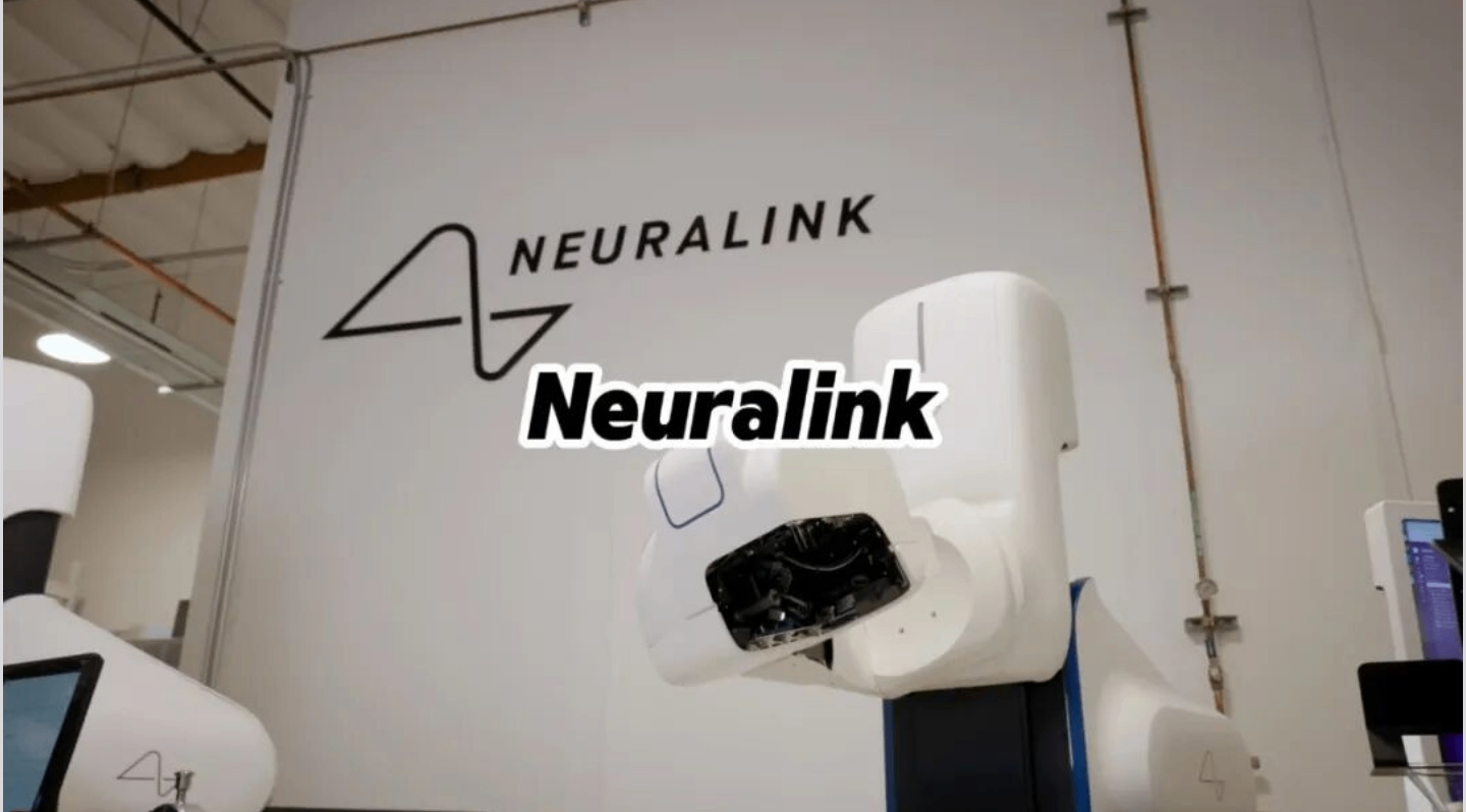
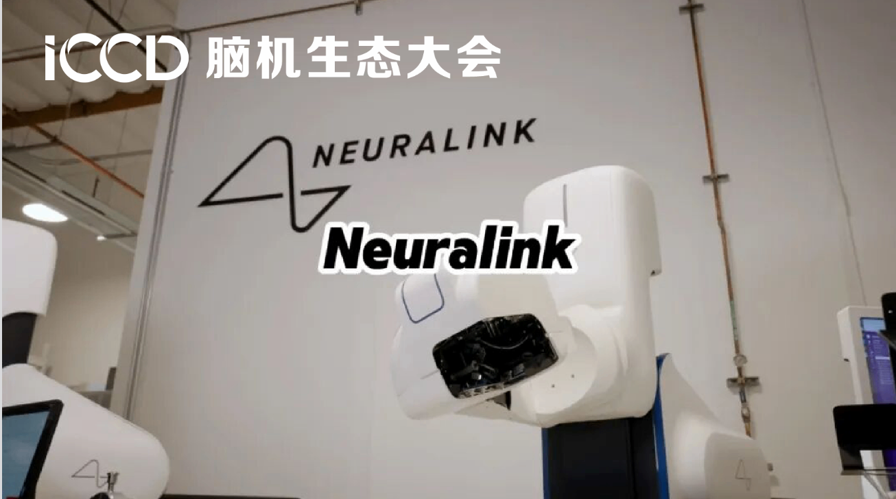
The R1 automated implantation surgical robot launched by Neuralink is one of the world’s few surgical platforms tailored specifically for Thread flexible electrodes that have advanced to the clinical stage. The intraoperative Optical Coherence Tomography (OCT)‑guided navigation system serves as the core sensing hardware of this automated workflow. Powered by this “optical vision plus precision robotics” combination, invasive BCI surgeries are breaking free from the constraints of individualized manual manipulation and evolving toward standardized, scalable clinical solutions.
Practical Bottlenecks: Conventional Electrode Implantation Solutions Struggle to Meet the Development Demands of High‑Density BCI
For a long time, neurosurgical electrode implantation has heavily relied on surgical microscopes and the individual experience of surgeons, revealing numerous unavoidable shortcomings in clinical practice. Conventional surgical microscopes only capture surface‑level views of the brain, making it impossible to pre‑identify numerous microvessels beneath the dura mater and within the superficial cerebral cortex. Even with high‑precision pre‑operative MRI scans, the outputs are merely static images. Following craniotomy, cerebrospinal fluid loss triggers brain‑tissue shift, creating inherent discrepancies between pre‑operative imaging and the real‑time intraoperative anatomical landscape, which limits its utility for real‑time risk mitigation.
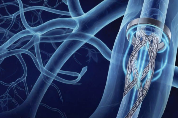
Looking at global peers, competing intracortical invasive solutions such as Blackrock and Paradromics still rely on manual surgical implantation by surgeons at this stage, without integrated real‑time imaging and automatic obstacle‑avoidance systems. On the other hand, intravascular‑interventional BCIs represented by Synchron’s Stentrode avoid craniotomy‑related trauma; however, constrained by their physical form factors, they face clear upper limits in channel count and information bandwidth. Against this industry landscape, automated implantation robots integrated with OCT‑guided navigation have become an indispensable building block for large‑scale clinical adoption of high‑density intracortical invasive brain‑computer interfaces.
Technology Implementation: How OCT Addresses Sensing Limitations of the Neuralink Implant Robot
Optical Coherence Tomography (OCT) can be figuratively understood as an “optical ultrasound system”. Based on the interferometric imaging principle of near‑infrared light, it generates tomographic three‑dimensional images by penetrating superficial tissues without contrast‑agent injection. Boasting three key features — ultra‑high resolution of 5‑20 μm, real‑time tomographic imaging, and label‑free non‑invasive detection — OCT is well‑suited for minimally invasive intracranial surgical scenarios. The complete optical module is compactly integrated into the robot end‑effector and forms a multimodal sensing network together with multiple high‑definition vision cameras.
1.Perform three‑dimensional mapping of cortical blood vessels to achieve dynamic obstacle avoidance.
Once the surgery commences, OCT rapidly scans the target surgical field and reconstructs the three‑dimensional cortical structure within a depth range of 1‑1.5 mm to precisely localize microvessels at the micrometer scale. Drawing on the imaging outputs, the AI‑powered path‑planning system generates an independent implantation trajectory for each Thread flexible electrode, ensuring sufficient safety margins between puncture sites and blood vessels. During continuous electrode advancement, OCT captures images uninterruptedly. Whenever physiological brain‑tissue movement causes path deviation, the robot swiftly corrects puncture coordinates to actively steer clear of hidden blood vessels, mitigating the risk of intracranial hemorrhage at its source.
2.Enable minimally‑invasive puncture across the dura mater and deliver precise control over puncture depth.
New‑generation technical solutions attempt to perform implantation by directly penetrating the dura mater, without requiring large‑scale dural incision, which can effectively reduce surgical trauma and the risk of infection. Nevertheless, the intact dura mater blocks conventional optical lenses, making it difficult to determine the actual distance from the dura mater to the cerebral cortex. With OCT tomographic imaging, the system can measure the thickness of multi‑layer tissues in real‑time, provide continuous feedback on needle‑tip position, and establish closed‑loop control to adjust puncture force and feed depth, preventing mechanical impact‑induced injury to cortical neurons by the needle tip.
3.Compensate for positioning errors introduced by physiological brain‑tissue motion.
Following craniotomy, cerebrospinal‑fluid loss and continuous physiological bodily movement trigger millimeter‑scale displacement of brain tissue. Capturing spatial tissue information at high frequency on an ongoing basis, OCT dynamically updates the robot’s spatial coordinate system and corrects puncture target points in real time. It offsets localization deviations inherent to static pre‑operative imaging and ensures consistent deployment of hundreds of electrodes into target functional brain regions.
At the hardware coordination layer, the R1 robot adopts a five‑axis linked motion platform with a repeat positioning accuracy at the 10‑micrometer level. An ultra‑fine puncture cannula grasps the polyimide‑based flexible Thread electrode. Upon reaching the target puncture site, the cannula releases the electrode and retracts in a controlled sequence. Early‑generation hardware required approximately 17 seconds to implant a single electrode. Following the introduction of real‑time OCT navigation and continuous iterative optimization of path‑planning algorithms, the implantation duration for one electrode has been reduced to 1.5 seconds. The overall multi‑electrode implantation workflow is greatly shortened. The long‑term goal is to complete full electrode deployment within one hour.
Clinical and Industrial Value: Automated Implantation System Beyond Surgical‑Accuracy Improvement
When Neuralink is widely discussed, most attention falls on the signal‑acquisition capabilities of its electrodes and chips, while the long‑term impacts brought by its surgical hardware are easily overlooked. This automated robot equipped with OCT‑guided navigation delivers value far beyond mere improvement in puncture precision.
First and foremost, it substantially improves surgical safety and lowers operational barriers. Leveraging the active OCT obstacle‑avoidance mechanism, the surgery becomes less reliant on surgeons’ hands‑on microsurgical experience. Standardized automated workflows reduce human‑operated errors. In the long run, this facilitates the roll‑out of invasive BCI implantation procedures across a broader range of medical institutions.
Secondly, it improves the long‑term service life of electrodes. A gentle and precise implantation approach mitigates acute brain‑tissue injury caused by puncture. It is expected to alleviate postoperative glial scar proliferation, slow signal attenuation of flexible electrodes, and extend the effective working cycle of implants. This also represents a pressing pain point to be addressed within the field of invasive BCI.
Most importantly, it lays the foundation for large‑scale commercialization. For invasive brain‑computer interfaces to move beyond clinical trials and deliver tangible benefits to patients suffering from neurological disorders such as amyotrophic lateral sclerosis (ALS) and high‑level paraplegia, stable, replicable and standardized surgical systems must be established. The integrated solution combining implantable electrodes, dedicated surgical robot and intraoperative real‑time imaging navigation forms Neuralink’s unique industrial‑chain moat that sets it apart from other BCI vendors. An electrode array alone can hardly constitute a complete clinical solution.
Real‑World Challenges and Medium‑to‑Long‑Term Evolution Pathways
That said, we should also take an objective view that the OCT‑guided implantation‑robot approach still presents numerous engineering challenges requiring continuous research and development.
For one thing, OCT light has a limited penetration depth and can only cover superficial cortical tissues. For targets located in deep‑brain regions, multi‑modal registration and fusion with pre‑operative MRI images become mandatory. For another, the full surgical platform features high integration, resulting in persistently high hardware costs. Device miniaturization and cost optimization represent unavoidable tasks for large‑scale popularization. Beyond that, biological issues including continuous micro‑motion control of brain tissue, post‑insertion retraction of flexible electrodes, and long‑term biocompatibility of electrodes demand coordinated optimization across multiple disciplines: imaging navigation, robotic control, and electrode materials.
In the medium‑to‑long term, technical iterations will most likely advance along several directions: advancing the fusion of OCT with fluorescence imaging and multispectral vision to build a multi‑level intraoperative sensing system; pushing forward full‑process robot automation to realize integrated operations including skull drilling, dural puncture and electrode implantation; optimizing supporting algorithms and exploring the prediction of postoperative tissue responses based on intraoperative images, so as to achieve truly personalized planning of implantation targets.
Implications for China’s Invasive Brain‑Computer Interface Track
Looking across China’s domestic BCI industry, most enterprises at the current stage focus on semi‑invasive ECoG approaches, including subdural and epidural solutions. Only a handful of teams are pursuing intracortical invasive directions. Projects capable of advancing the full system covering flexible electrodes, automated implantation devices and intraoperative optical navigation in parallel are even scarcer.
Neuralink’s development path offers a clear reference for domestic practitioners. Competition in high‑density invasive brain‑computer interfaces cannot be confined to electrodes and chips alone. Surgical implantation hardware and intraoperative real‑time sensing‑navigation systems hold equally important strategic status. Even with high‑performance flexible electrode arrays, without a safe, stable automated implantation platform featuring autonomous obstacle‑avoidance capability, electrodes can hardly be implanted into the human brain smoothly and long‑term. Consequently, the performance ceiling of the hardware itself cannot be brought into play.
It is often figuratively compared that invasive BCI implantation is akin to “laying wires on tofu”, a vivid metaphor illustrating the extreme difficulty of this task. The R1 surgical implantation robot equipped with OCT optical navigation uses optical imaging as an intracranial detection radar and performs micrometer‑scale operations via precision machinery, clearing the critical barrier for the safe implantation of flexible electrodes.
Light penetrates intracranial tissues to reveal intricate vascular networks; intelligent machinery carries out precise operations to build a communication bridge between the human brain and machines. With the continuous coordinated evolution of intraoperative imaging, robotic control and flexible neural electrode technologies, minimally‑invasive automated surgical solutions represented by the OCT‑guided implantation robot will keep driving invasive brain‑computer interfaces out of the laboratory. In the future, such technologies are expected to deliver new therapeutic options for large populations suffering from impaired neurological function, progressively advancing human‑machine interaction into a new era.